登录

- APP
中国粉体网欢迎您�?/div>
- 粉享這�/a>
- 188188188b.com�𱦲�
微信

 关注微信公众叶�/span>
- 中国粉体罐�/a>
 移动�?/p>
 m.cnpowder.com.cn
登录
微信

 关注微信公众叶�/span>
![]() 移动�?/p>
移动�?/p>
 m.cnpowder.com.cn

 留言询价
留言询价

虚拟号将�180秒后失效
使用微信扫码拨号
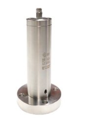
Navitar 深紫�?DUV)光学系统
NanoVue 248nm 4x 深紫外变焦系� ( 光学检� 0.1um)
提供工业级深紫外变焦解决方案
针对进一步扩大光学分辨率的需求,Navitar开发出NanoVue 248nm 4X深紫外变焦镜头,用于光学检测。NanoVue光学系统工作在深紫外(DUV)波段,用于检测和分析关键的缺陷。相对于传统的可见光显微镜,NanoVue成倍地提高了分辨率的极限、�/span>
90nm分辨玆�/span>
放大倍率75X - 300X� 配合100X 物镜
0.15 to 0.037 mm FOV 对应 2/3" 格式传感器和100X物镜
采用深紫外物镜,检测尺度可�?.1 um
工业级深紫外变焦解决方案
应用
? UV reticle & photomask inspection (UV 投影和掩模检�?
? Thin film measurement ( 薄膜测量)
? Wafer inspection ( 晶圆检�?
? CD metrology ( 关键尺寸测量)
? Failure analysis ( 失效分析)
? Process control ( 过程控制)
? Protein crystal location ( 蛋白质晶� 率选和� 定位)
? Pharmaceutical quality control ( 药物质量控制)
? Contaminant analysis ( 杂质分析)
? Cellular imaging & DNA analysis ( 细胞成像和DNA 分析)
NanoVue变焦镜头是理想的新产品开发和集成DUV仪器的工具,具有更高的光通量,允许客户快速开发新的、灵活的自动化仪器,具备功能丰富、成本更低、体积更小等特点、�/span>
以下是利用深紫外(DUV) 光学系统� 蛋白晶体 (crystalline of a protein) 进行筛选的应用
案例� 请参考� -- Picking out proteins with UV.
Picking out proteins with UV
By Mike May
In the late 1990s� Vu Tran� president of Korima (Carson� CA)� and Gil Ravich� president of Ravich Research (Lawndale� CA)� developed a microscope that could image samples under ultraviolet (UV) light. They developed this scope to “see� down to about 0.2 ��m to examine semiconductors. When electronics makers started using nitrides� though� UV couldn?t go through
them. That turned Tran and Ravich?s scope into a fossil. “I was very frustrated,� says Tran� “and I put the microscope in my garage and said� ?Forget it!? “�/span>
Then� in the summer of 2003� Tran and Ravich?s luck changed. Someone from a pharmaceutical company–they won?t say which one–approached them about using their scope to image proteins. By adding two filters� the pair made their UV microscope focus on light at 280 nm� where crystalline proteins fluoresce and are thus distinguishable from salt crystals. Making this distinction is a key step in deciding whether to move a protein to x-ray crystallography to find its structure� which helps pharmaceutical scientists understand a disease target or refine a drug. Unfortunately� the pharmaceutical company and Korima couldn?t agree about ownership of intellectual property� so Tran and Ravich put away their scope once more.
Figure 1. Results of work done to make a crystalline form of a protein can be difficult to see under visible light ()� but protein crystals become quite clear under ultraviolet light at 280 nm (bottom). Moreover� at 280 nm� protein crystals can be distinguished from salts. (Images courtesy of Korima)
Eventually� Korima?s scope� the PRS-1000 Protein Review Station� did come on the market–a market that is getting bigger in terms of customers and vendors.
Ultraviolet vision
Although various nomenclatures define UV?s span differently� it is said to range from 10 or so nanometers to 400. “One advantage to using UV microscopy is that it allows you to image features that would not normally be seen with a standard visible-range microscope,� says Paul Martin� president of CRAIC Technologies (San Dimas� Calif.). The shorter wavelengths of UV reveal things that visible light cannot.
In many situations� though� scientists want an instrument that works over a range of wavelengths. Consequently� CRAIC developed the UVM-1 Ultraviolet Microscope� which works in UV� visible� and near-infrared (NIR) regions. “It was a real challenge to achieve high image quality while using the same optics over such a broad spectral region,� says Martin.
Given growing competition� however� no one reveals the precise details behind any optical device. As Martin says� it requires “experience� advanced optical design� and extensive experimentation.“�/span>
Some companies apply such experience to specific pieces� like the NanoVue 248 nm 4X Deep UV Zoom from Navitar (Rochester� NY). William Bridson� director of research and development at Navitar� says that this lens “is being used in a number of common biological applications where specimens are transparent in the visible region� but absorb light in the ultraviolet spectrum.� Beyond looking for protein crystals� says Bridson� this lens can be used for “pharmaceutical quality control� contaminant analysis� cellular imaging� and DNA analysis.“�/span>
UV microscopy also requires the right illumination. While Rapp Optoelectronics (Hamburg� Germany) makes UV microscopes� it also makes light sources. “We use white light� and then filters and optics optimize the wavelength,� says Gert Rapp� general manager. If someone needs high-power light at� say� 220 nm� Rapp uses pulsed xenon light as the source. In short� though� different applications need different features. As an example� Rapp notes that “DNA absorbs strongly at around 260 nm� so you can measure it directly.“�/span>
Rapp Optoelectronics will even modify a customer?s existing microscope for UV applications. “We?ve done that on a variety of microscopes,� Rapp says.
Digging even deeper
To look even closer at samples� some scientists turn to x-rays. Conventional x-rays–because of their high energy–go right through a sample and get detected on a photographic plate. Soft x-rays� though� are lower in energy� and they get absorbed. “Soft x-rays use diffraction optics. You can make lens and mirrors and nanostructures with features about the size of the wavelengths,� explains David T. Attwood� professor in residence in electrical engineering and computer science at the University of California at Berkeley and director of the Center for X-Ray Optics at the Lawrence Berkeley National Laboratory.
These wavelengths for soft x-rays go down to about 20 nm� at the shortest range of what some scientists still call UV. “At 20 nm,� says Attwood� “all materials are absorptive there.� So soft x-rays provide natural contrast with biological materials. Compared with looking at protein crystals at 280 nm� a soft-x-ray system increases the resolution by a factor of more than 10.
Figure 2. Ultraviolet-viewing setups require specialized pieces� such as the NanoVue 248 nm 4X Deep UV Zoom used in this microscope. (Image courtesy of Navitar).
Consequently� soft-x-ray microscopy can be used to image a variety of subcellular features. “In the cylasm,� says Attwood� “you would see the vesicles� and you can measure their variant absorption.“�/span>
Still� soft x-rays create some obstacles. For example� a sample must be fixed� so you can?t see dynamic events. Nonetheless� Attwood points out that soft-x-ray microscopy is faster than electron microscopy� while providing similar resolution. “You can look at lots of samples and do statistics,� he says. “You can also do so with three-dimensional imaging.� A researcher� for
example� could look at the distribution of proteins and then knock out a gene and see if the distribution changes.
In the June 2008� Journal of Structural Biology� Dilworth Y. Parkinson of the Lawrence Berkeley National Laboratory and colleagues describe using soft x-rays to create three-dimensional images of schizosaccharomyces pombe–or yeast–cells. As Dilworth and colleagues write: “In addition to imaging intact cells� soft-x-ray tomography has the advantage of not requiring the use of any staining or fixation protocols–cells are simply transferred from their growth environment to a sample holder and immediately cryofixed. In this way the cells can be imaged in a near native state.
“Soft-x-ray tomography is also capable of imaging relatively large numbers of cells in a short period of time� and is therefore atechnique that has the potential to produce information on organelle morphology from statistically significant numbers of cells.“�/span>
As with many advances in microscopy� UV and its near neighbors give scientists a closer look at biology. Moreover� researchers are only beginning to see what this technology can reveal
暂无数据�
 Navitar深紫�?DUV)光学系统的工作原理介绍?Navitar深紫�?DUV)光学系统的使用方法?Navitar深紫�?DUV)光学系统多少钱一台?Navitar深紫�?DUV)光学系统使用的注意事顸�/li>Navitar深紫�?DUV)光学系统的说明书有吗>�/li>Navitar深紫�?DUV)光学系统的操作规程有吗?Navitar深紫�?DUV)光学系统的报价含票含运费吗?Navitar深紫�?DUV)光学系统有现货吗>�/li>Navitar深紫�?DUV)光学系统包安装吗>�/li>
 手机版:
手机版: