虚拟号将 180 秒后失效
使用微信扫码拨号
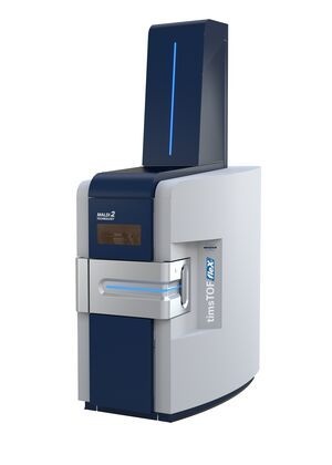
布鲁克于ASMS2019上推出高度创新的新型质谱产品timsTOF fleX?质谱仪,包括适配于ESI timsTOF Pro?质谱的软件可切换的MALDI源。这种新型ESI/MALDI组合功能,可在单台质谱上实现空间定位组学SpatialOMx?。timsTOF fleX配备布鲁克独有的10 kHz SmartBeam? 3D激光源,具有真正的像素保真度,可在高空间分辨率下进行快速、无标记的MALDI成像,同时完全保持timsTOF Pro在ESI模式?***?D蛋白质组学和表型组学的灵敏度、/span>
利用这种独特的SpatialOMx方法,研究人员可以深入洞悉MALDI成像组织中的空间分子分布,从而指?D组学分子表达研究,例如蛋白质、低水平癌抗原肽、脂质、聚糖、代谢物或那些用传统染色或标记技术无法观察到的分子。在MALDI指导下的SpatialOMx方法中,使用者可用随后由ESI-TIMS/PASEF的dda(数据依赖采集),或者dia(非数据依赖采集?D蛋白质组学或4D脂质组学/代谢组学的数据,来开展细胞亚群的特异性靶向研究。两者现在都可以在同一台强大的仪器上进行,即timsTOF fleX,具有推进单细胞生物学研究所需的超高灵敏度,成为用RNA测序(RNA-seq)进行单细胞转录组学研究?*补充、/span>
The microenvironment of cancers contains a combination of healthy cells tumor cells connective tissue blood vessels and inflammation in different ratios at different time points. Each of these components will have their own unique-molecular signature of compounds. Researchers who study disease states rely heavily on interpreting tissue pathology and creating these maps within the context of biomolecules bridges the gap between traditional OMICS and understanding disease.
Cancer cells and other disease states have significant genetic and epigenetic modifications that influence the genomic expression cascade. Whether you’re looking at the proteome lipidome or metabolome the spatial distribution of compounds contains valuable Information for understanding your sample. If certain compounds are highly spatially concentrated or if molecules are co-distributed on specific structures this vital information is missing when only examining homogenized samples. OMICS based biomarker discovery can be limited without the benefit of spatial information to add important contextual clues as tissue-level communications networks are integral to cancer growth.
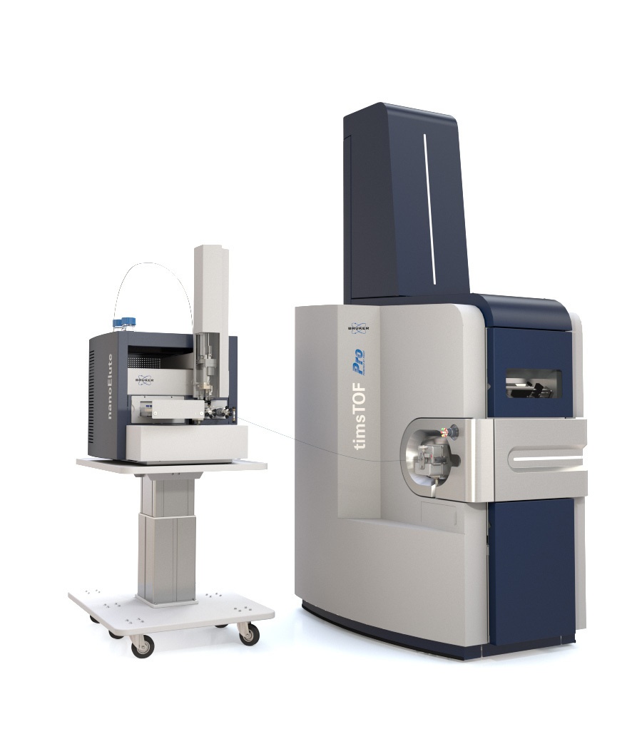
The timsTOF fleX is a versatile instrument for probing the molecular content of your sample. Built on Bruker’s pioneeringtimsTOF Proplatform the timsTOF fleX is a fully functional high speed high sensitivity ESI instrument for all your X-Omics analyses combined with a high spatial resolution MALDI source and stage specifically designed for resolving molecular distributions and bringing a spatial dimension toOMICS analyses. Transform proteomics analysis into spatial proteomics lipidomics into spatial lipidomics and metabolomics into spatial metabolomics.
Mapping complex molecular networks exposes intricate systems of interacting molecules within intracellular networks. Using SpatialOMx to create insightful atlases of communication mechanisms means learning more about the way cells communicate with each other to support change:
how cells interact with the local microenvironment to formulate promotion and mediation processes
how molecular networks of specific cells interact to perform specific biological functions
what effect epigenetic modifications have within these networks.
The timsTOF fleX the flexibility to switch between spatial metabolomics spatial lipidomics or spatial proteomics with ease.
Match PASEF powered LC-MS/MS identification with label-free spatial localization to decode the molecular make up of your sample
Built on the standard for shotgun proteomics the timsTOF fleX combines best in class 4D X-Omics with Bruker’s cutting edge MALDI Imaging technology including SmartBeam 3D laser optics for fast measurement all in one platform. A dual source instrument ideal for SpatialOMx timsTOF fleX conducts robust ESI measurements and spatially resolves a wide range of molecules directly from tissue using one platform. No single instrument has before provided access to both essential capabilities for the most advanced OMICS researchers.
SpatialOMx demands PASEF enabled LC-MS/MS for deep 4D OMICS combined with the ability to create molecular images of tissue sections with MALDI Imaging. To do this under one platform Bruker engineers started with the award winning TIMS dual-stage ion funnel and incorporated target stage and laser elements from the rapifleX axial TOF instrument. During operation software activation of the SmartBeam 3D laser is the only change in the source region. No complicated changeover means making zero compromises in productivity and the ability to move effortlessly from world class OMICS identification and quantification workflows to creating high definition molecular maps of tissue sections to see what matters most.
Since ions produced by MALDI and ESI travel the same path to the detector from the source MALDI workflows can take advantage of the most advanced features found in the timsTOF Pro including TIMS ion mobility separation based on the collisional cross section of detected molecules and high-speed high-sensitivity PASEF fragmentation. Tuning and calibration can be performed in ESI mode and used for the MALDI experiment for additional ease of optimization.
MALDI Imaging is a label-free tool for examining the distribution of molecules directly from thin tissue sections. Applicable for examining a wide range of analytes including metabolites lipids or glycans and seamlessly compatible with microscopy workflows MALDI Imaging can identify regionally specific compound distributions for SpatialOMx analyses. Use Bruker’s IntelliSlides for automated MALDI Imaging and SpatialOMx workflows on the timsTOF flex.
Besides the ability to generate high definition “True Pixel MALDI images the SmartBeam 3D laser source provides rock solid laser guided targeting down to 10 M with Bruker’s new zoom mode technology. Users now have the flexibility to adjust their laser spot size and spacing to accommodate a wider range of granularity depending on biological features of interest.
Combining X-Omics performance with high MALDI sensitivity
Whether it’s proteomics lipidomics glycomics or metabolomics the timsTOF fleX is the ideal platform for deep SpatialOMx analyses. Use patented SmartBeam? 3D technology to conduct fast label-free MALDI Imaging for mapping the distribution of molecules in your sample and identify regions of interest for more specific analyses. ApplyPASEF powered LC-MS/MSfor the highest level of identifications and for greatest confidence in your results.
Combining the power of trapped ion mobility spectrometry (TIMS) andParallel Accumulation Serial Fragmentation (PASEF) the timsTOF fleX can deliver quantitative information from smaller amounts of highly complex mixtures. Using less than 200 ng sample load and a 90 min gradient length more than 5400 protein groups can be identified from a typical human cell line lysate. The high sensitivity reduces sample preparation costs and decreases time lost to maintenance.
With existing protocols for lipidomics metabolomics or glycomics MALDI Imaging spectra can be used as a molecular fingerprint for specific regions or to investigate how compounds are localized across an entire sample for biomarker discovery. This can also be used for investigation of temporal changes quantification and correlation of compounds to tissue states. The timsTOF fleX allows for fast high mass resolution MALDI imaging with the SmartBeam 3D and specialized MALDI source and stage a high spatial resolution measurement of lipids from a rat brain section consisting of 370?00 pixels takes less than 5 hours.
Traditional workflows that sample small tissue sections (<100 M) provide deep molecular details for a variety of chemical classes including RNA/DNA proteins glycans lipids and metabolites. However processing times are long and tissue excise locations are determined with optical images that lack molecular signal information. SpatialOMx on the timsTOF fleX rapidly provides a high definition label-free overview that merges chemical information with vital morphological context. Choose your regions of interest manually or use SCiLS enabled automated segmentation to know exactly where your extraction will have the highest impact.
Using Bruker’s industry leading software it is now possible to annotate target molecules directly from tissue. Simply load your data into SCiLS Lab define regions of interest and export the peak lists to MetaboScape. Annotate your peaks using databases or compound lists from LC-MS/MS analyses then review and export your annotations back to SCiLS for visualization. From SCiLS you can then visualize results with pathways and familiar nomenclature rather than mass labels to reduce the time from data to results.
南卡罗来纳医科大学蛋白质组学中心主任Richard Drake教授表示:“使用新型timsTOF fleX系统运行的样本,其数据在空间分辨率和聚糖覆盖深度方面?***的。聚糖已成为组织和血清中潜在的临床标志物,用以监测身体的免疫状态,正常或非正常的衰老,timsTOF fleX的独特功能将大大加速该研究进程 timsTOF fleX使得我们可将为癌症和免疫疗法的组织和生物流体分析开发的方法汇集在一个平台上。我可以看到这款仪器在许多研究领域中的无限应用,比如快速聚糖组织成像和生物流体4D组学分析。“/span>
布鲁克道尔顿执行副总裁Rohan Thakur博士补充说:“大多数组织蛋白质组学研究融合了来自不同亚群的细胞,其不期望的平均效应掩盖了许多重要的生物学和病理生物学信息。在timsTOF fleX上,MALDI引导的SpatialOMx方法能够在空间上对指定的组织区域进行分析,从而允许随后的4D蛋白质组学选择性地对细胞类型的亚群进行靶向分析。功能强大、具有超高灵敏度的timsTOF fleX,在同一仪器上还提供nanoLC-TIMS-MS/MS,从而弥合了分子组织成像和体液分析间的鸿沟。这种独特的组合将使timsTOF fleX成为宝贵的研究工具,用于空间分辨?D蛋白质组学和表型组学,并推动单细胞生物学、药物蛋白质组学和大细胞数量或患者队列的转化4D交叉组学的工作流程。“/span>
Helmholtz药物研究所(HIPS)助理教授Daniel Krug博士评论说:“我们在药物研究和空间代谢组学领域使用质谱技术;尤其是分析新型天然产物的粘细菌次级代谢组。我们实验室多年来一直使用布鲁克的仪器和软件,特别是SCiLS Lab和MetaboScape;新分子成像工作流程集成了这两种软件解决方案,极大地简化并加速了整个MALDI成像流程。该流程将离子图像转换为分子图像,并提高了注释分子式的置信度。“/span>
暂无数据